
Page 77 - 《应用声学》2021年第1期
P. 77
第 40 卷 第 1 期 刘强等: 磁纳米粒子介导的靶向剪切波弹性成像 73
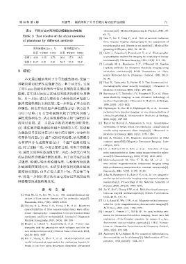
表 2 不同方法对两种剪切模量仿体的检测 vibrometry[J]. Medical Engineering & Physics, 2017, 39:
66–72.
Table 2 Test results of the shear modulus
[5] Guo Y, Lin H, Dong C, et al. Role of acoustic radiation
of phantoms by different methods
force impulse imaging elastography in the assessment of
steatohepatitis and fibrosis in rat models[J]. Medical En-
剪切波速度/(m·s −1 ) 剪切模量/kPa gineering & Physics, 2018, 59: 30–35.
仿真 TSWE AFRI 仿真 TSWE AFRI [6] Ophir J, Cespedes I, Ponnekanti H, et al. Elastography:
仿体 1 4.01 3.65 3.75 20.9 17.3 18.3 a quantitative method for imaging the elasticity of biolog-
ical tissues[J]. Ultrason Imaging, 1991, 13(2): 111–134.
仿体 2 6.37 6.31 6.61 52.7 51.8 56.8
[7] Lubinski M A, Emelianov S Y, O’Donnell M. Speckle
tracking methods for ultrasonic elasticity imaging using
3 结论 short-time correlation[J]. IEEE Transactions on Ultra-
sonics Ferroelectrics & Frequency Control, 1999, 46(1):
本文通过磁纳米粒子介导的磁致振动,发展一 82–96.
种靶向剪切波弹性成像新方法。基于该方法,实现 [8] Hoyt K, Castaneda B, Parker K J. Two-dimensional so-
noelastographic shear velocity imaging[J]. Ultrasound in
了对 3 mm 的磁性纳米粒子标记区域的高灵敏成像 Medicine & Biology, 2008, 34(2): 276–288.
检测,并实现对该标定区域周围组织的弹性信息获 [9] Sarvazyan A P, Rudenko O V, Swanson S D, et al. Shear
取。另一方面,通过与理论仿真和传统的声辐射力 wave elasticity imaging: a new ultrasonic technology of
medical diagnostics[J]. Ultrasound in Medicine & Biology,
脉冲成像检测结果相比较,进一步验证了本方法的 1998, 24(9): 1419–1435
准确性。相比传统的超声弹性成像方法,该方法具 [10] Nightingale K, Soo M S, Nightingale R, et al. Acoustic
有以下优势:(1) 有望将磁纳米粒子的靶向识别与 radiation force impulse imaging: in vivo demonstration of
clinical feasibility[J]. Ultrasound in Medicine & Biology,
弹性成像相结合,从而实现肿瘤标志物与弹性信息 2002, 28(2): 227–235.
的同时获取,进一步提高诊断的灵敏性和特异性; [11] Tanter M, Bercoff J, Athanasiou A, et al. Quantitative
(2) 通过脉冲磁场激励和超声探测的方式,有望解 assessment of breast lesion viscoelasticity: initial clinical
results using supersonic shear imaging[J]. Ultrasound in
决激励信号在活体复杂环境中的穿透性、安全性和 Medicine & Biology, 2008, 34(9): 1373–1386.
特异性等问题;(3) 超声灰阶结构成像、纳米颗粒分 [12] Issa B, Obaidat I M. Magnetic nanoparticles as MRI
布和弹性分布成像有望在同一个超声检测系统完 contrast agents[M]//Magnetic Resonance Imaging. Inte-
chOpen, 2019.
成,时空坐标一致,不需要配准过程,有利于图像融
[13] Oh J, Feldman M D, Kim J, et al. Detection of mag-
合,进而实现多模态成像分析。因此,本研究有望为 netic nanoparticles in tissue using magneto-motive ultra-
疾病的精准诊断提供新的思路。由于该方法的成像 sound[J]. Nanotechnology, 2006, 17(16): 4183–4190.
[14] Mehrmohammadi M, Shin T H, Qu M, et al. In
灵敏度、检测信噪比和检测深度,与检测部位的脉
vivo pulsed magneto-motive ultrasound imaging using
冲磁通密度密切相关,本研究中所采用的脉冲磁通 high-performance magnetoactive contrast nanoagents[J].
密度相对较弱,仍具有很大提升空间。在后续工作 Nanoscale, 2013, 5(22): 11179–11186.
[15] John R, Rezaeipoor R, Adie S G, et al. In vivo magneto-
中,将进一步探讨该方法对亚毫米标记区域及动物
motive optical molecular imaging using targeted magnetic
肿瘤模型的成像检测效果。 nanoprobes[J]. Proceedings of the National Academy of
Science, 2010, 107(18): 8085–8090.
参 考 文 献 [16] Jin Y, Jia C, Huang S W, et al. Multifunctional nanopar-
ticles as coupled contrast agents[J]. Nature Communica-
[1] Tian M, Li Y, Liu W, et al. The nanomechanical sig-
tions, 2010, 1(4): 41.
nature of liver cancer tissues and its molecular origin[J].
[17] Li J, Arnal B, Wei C W, et al. Magneto-optical nanopar-
Nanoscale, 2015, 7(30): 12998–13010.
ticles for cyclic magnetomotive photoacoustic imaging[J].
[2] Choong K L, Wong Y H, Yeong C H, et al. Elasticity
ACS Nano, 2015, 9(2): 1964–1976.
characterization of liver cancers using shear wave ultra-
[18] Loupas T, Powers J T, Gill R W. An axial velocity esti-
sound elastography: comparison between hepatocellular
mator for ultrasound blood flow imaging, based on a full
carcinoma and liver metastasis[J]. Journal of Diagnostic
evaluation of the Doppler equation by means of a two-
Medical Sonography, 2017, 33(6): 481–488.
dimensional autocorrelation approach[J]. IEEE Transac-
[3] Riegler J, Labyed Y, Rosenzweig S, et al. Tumor elas-
tography and its association with collagen and the tu- tions on Ultrasonics Ferroelectrics & Frequency Control,
1995, 42(4): 672–688.
mor microenvironment[J]. Clinical Cancer Research, 2018,
[19] Anderssen R S, Hegland M. For numerical differentiation,
24(18): 4455–4467.
[4] Lin H, Zhang X, Shen Y, et al. Model-dependent and dimensionality can be a blessing! [J]. Mathematics of Com-
model-independent approaches for evaluating hepatic fi- putation, 1999, 68(227): 1121–1141.
brosis in rat liver using shearwave dispersion ultrasound